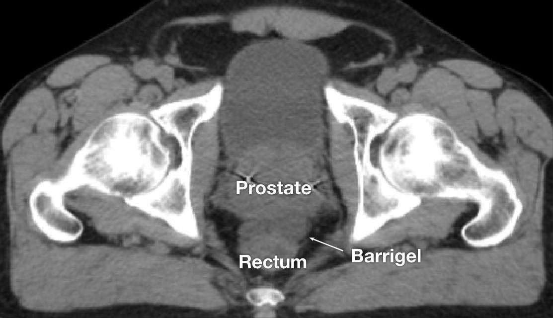
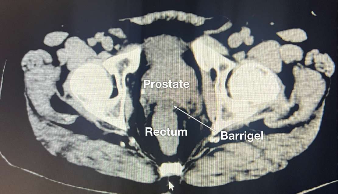
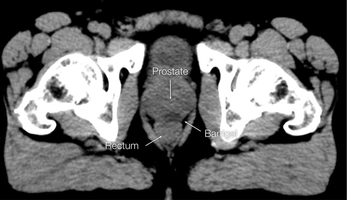
CT image courtesy of Gregory Bell, MD Interventional Radiologist; Texas, United States
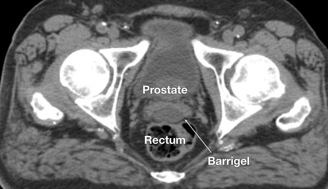
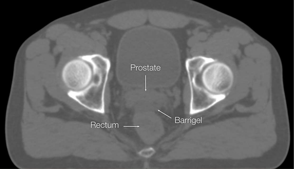
CT image courtesy of Suraj Singh, MD Radiation Oncologist, Colorado, United States
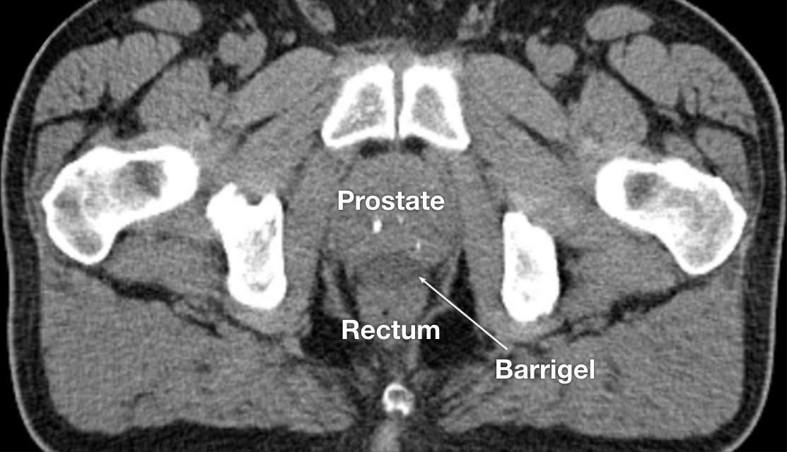
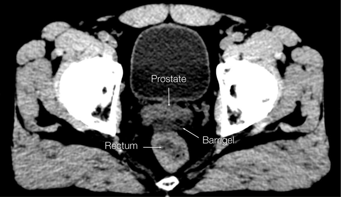
CT image courtesy of Suraj Singh, MD Radiation Oncologist, Colorado, United States
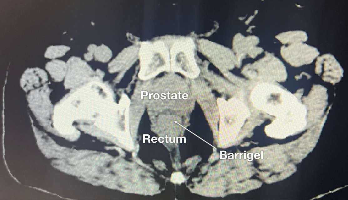
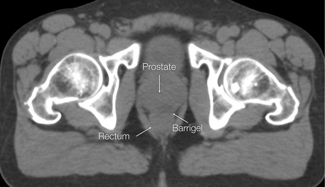
CT image courtesy of Suraj Singh, MD Radiation Oncologist, Colorado, United States
OPTIMIZING CT IMAGES
OPTIMIZING CT IMAGES
Barrigel is visible on CT. Window and level optimization can help bring out subtle density and texture differences, and can often enhance the Barrigel border contrast. Refer to the Barrigel CT Visualization Guide for detailed instructions.
Before (left) and after (right) adjusting window & level settings to enhance contrast and texture CT images sourced from the Barrigel Pivotal Trial
MRI IMAGES
MRI IMAGES
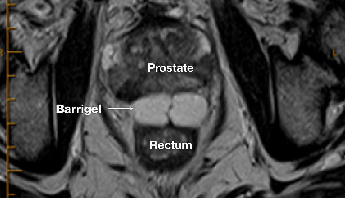
MR image courtesy of Prof Michael Chao, MBBS (Hons), FRANZCR, AFRACMA, DMedSc, Radiation Oncologist; Victoria, Australia
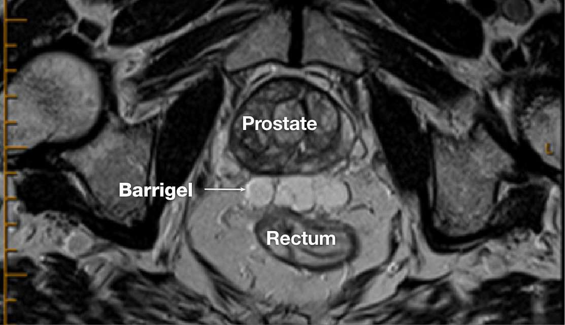
MR image courtesy of Prof Michael Chao, MBBS (Hons), FRANZCR, AFRACMA, DMedSc, Radiation Oncologist; Victoria, Australia
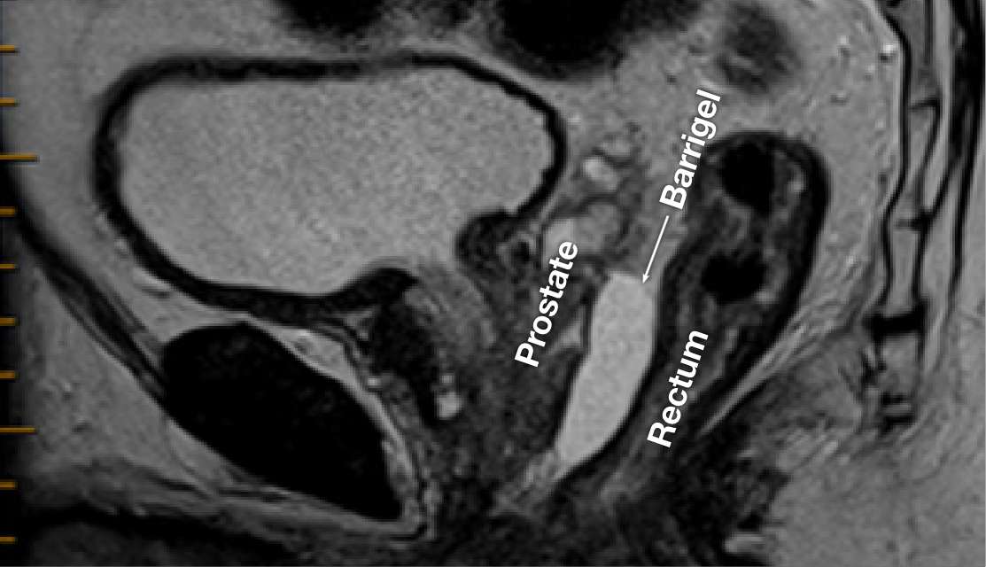
MR image courtesy of Prof Michael Chao, MBBS (Hons), FRANZCR, AFRACMA, DMedSc, Radiation Oncologist; Victoria, Australia